

Services
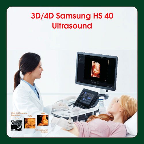
4D Color Ultrasound ( Fetal – Renal – Carotid – Peripheral Vessels )
A Doppler ultrasound likewise called a Color Doppler test is a non-invasive test that can be utilized to estimate your blood course through veins. It assists specialists with assessing blood flow through significant supply routes and veins, like those of the arms, legs, and neck. It can show hindered or diminished progression of blood through slender regions in the significant supply routes of the neck that could cause a stroke. It additionally can uncover blood clusters in leg veins (profound vein apoplexy, or DVT) that could loosen up and block the bloodstream to the lungs (pneumonic embolism). During pregnancy, Doppler ultrasound might be utilized to see the bloodstream in an unborn child (embryo) to really take a look at the soundness of the fetus. shading doppler-test.
What to Know About Test Results?
For a stomach ultrasound, you’ll rest and a specialist will put a specific gel on your paunch. These aides convey the sound waves. Then, at that point, the specialist will hold a test against your paunch and move it around to get a picture.
As long as 10 weeks (Early pregnancy)
Quantities of incipient organisms (Pregnancies) or various sacs.
Cardiovascular movement (pulses) and reasonability.
Regularities of the sac.How is the help for pregnancy?
To pass judgment on the fetus evacuation process.
Rundown of Common Color Doppler test techniques:
• Abdominal
• Carotid
• Gravid Uterus
• Renal
• Single Limb Doppler (Arterial and Venous)
• Dual Limb Doppler (Arterial and Venous)
A renal ultrasound is a secured and effortless test that utilizes sound waves to take pictures of the kidneys, ureters, and bladder. The kidneys are a couple of bean-molded organs situated rearward of the stomach hole, simply over the midsection. They eliminate byproducts from the blood and produce pee.
Transvaginal ultrasound
A transvaginal ultrasound utilizes a test that is embedded straightforwardly into the vagina. It is performed in the specialist’s office like a pelvic examination. This sort of test is most ordinarily utilized in the early long stretches of pregnancy to preclude suspected issues or to survey the gestational age of the undeveloped organism. In early pregnancy, this examination can give more precise data than a transabdominal examination.
High-Resolution Ultrasound Musculoskeletal
High-goal ultrasound is an amazingly helpful and versatile method for examining the musculoskeletal system. It has the benefit of being readily accessible and reasonable, simultaneously giving profoundly point-by-point data with respect to a variety of clinically important constructions of the musculoskeletal system, including tendons, muscles, nerves, ligaments, joints, and bone.
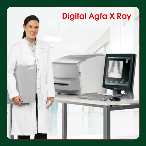 Digital X-ray replaces the utilization of movie or figured radiography (CR) plates with a direct advanced exchange of X-ray pictures into the PACS. Digital radiography (DR) is the immediate transformation of sent X-ray photons into a computerized picture utilizing a variety of strong state locators like undefined selenium or silicon, with computer processing and display of the picture. Least Radiation Dose/Maximum Diagnostic Accuracy 30% Dose Reduction/2.5x Higher DQE. Exceptional Patient Throughput and Fast Workflow. High DQE/MTF Value. DR X-Rays systems are utilized for both fixed-base room installations and mobile DR, or compact DR, Systems that are wheeled between spaces for imaging tests. Here is a connection to the convenient DR Systems product comparison diagram. Here is a connection to the DR Systems outline that incorporates fixed room-based Systems and retrofit systems to replace film and CR.
The improvement of computed radiography in the course of recent many years has changed radiological imaging. The radiology divisions in the 21st century will appear to be extremely unique from those in the previous period. In this survey, the improvement of digital radiography is given a portrayal of its different structures and a correlation with screen-film radiography.
Watchwords: digital radiography, figured radiography, demonstrative imaging
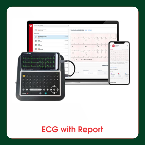
An electrocardiogram is a basic, easy test that measures your heart’s electrical movement. It’s otherwise called an ECG or EKG. Every heartbeat is set off by an electrical sign that starts at the most elevated mark of your heart and goes to the base. Heart issues consistently impact the electrical development of your heart. Your primary care physician might suggest an EKG in case you’re encountering side effects or signs that might recommend a heart issue, including:
- pain in your chest
- trouble in breathing
- feeling drained or weak
- beating, hustling, or rippling of your heart
- an inclination that your heart is thumping unevenly
- location of surprising sounds when your primary care physician pays attention to your heart
- An EKG will assist your primary care physician with deciding the reason for your indications alongside what kind of treatment may be important.
An ECG (electrocardiogram) records the electrical activity of your heart very still. It gives data about your pulse and musicality and shows assuming there is a growth of the heart because of (hypertension) or proof of a past coronary episode (myocardial localized necrosis). In any case, it doesn’t show whether you have asymptomatic blockages in your heart supply routes or foresee your danger of a future respiratory failure. The resting ECG is unique in relation to pressure or exercise ECG or cardiovascular imaging test.
Also known as echocardiography, 2D Echo utilizes ultrasound waves to produce real-time images of your heart. It allows healthcare professionals to examine your heart’s structure, assess its pumping function, and detect any abnormalities or conditions.
Using state-of-the-art technology, a skilled technician will place a probe on your chest, transmitting sound waves that create detailed images of your heart on a monitor. These images help doctors evaluate your heart’s valves, chambers, and blood flow patterns, helping to diagnose and monitor various cardiovascular conditions, such as heart attacks, valve diseases, and congenital heart defects.
With its ability to provide accurate and immediate results, 2D Echo has become an indispensable tool in diagnosing and treating heart conditions. Whether you are experiencing symptoms or simply want a comprehensive cardiovascular assessment, 2D Echo offers a safe and reliable diagnostic solution. So, let’s dive deeper into the world of 2D Echo and discover how it can help you take control of your heart health.
The 2D Echo plays a crucial role in diagnosing and monitoring a wide range of heart conditions. By providing detailed images of the heart’s structures and functions, it allows healthcare professionals to detect abnormalities, assess the severity of conditions, and guide treatment decisions. Let’s explore the importance of a 2D Echo in diagnosing various heart conditions.
1. Identifying structural abnormalities
A 2D Echo can detect structural abnormalities such as congenital heart defects, valve diseases, and enlarged chambers. It allows healthcare professionals to visualize the heart’s anatomy and identify any malformations or irregularities. This information is essential for diagnosing conditions present at birth or those that develop over time, enabling early intervention and appropriate treatment.2. Assessing heart function
Assessing the heart’s function is a key aspect of diagnosing heart conditions. A 2D Echo provides valuable information about the pumping efficiency, wall motion, and contractility of the heart. By evaluating these parameters, healthcare professionals can determine if the heart is functioning properly or if there are any abnormalities affecting its ability to pump blood effectively. This information is critical for diagnosing conditions such as heart failure, cardiomyopathy, and myocardial infarction.3. Evaluating valve function
The heart’s valves play a crucial role in maintaining proper blood flow through the chambers. A 2D Echo allows healthcare professionals to assess the function and integrity of the heart valves, detecting any abnormalities such as valve stenosis (narrowing) or regurgitation (leaking). This information helps diagnose and monitor valve diseases, guiding treatment decisions and interventions when necessary.4. Detecting blood clots and tumors
A 2D Echo can also detect the presence of blood clots or tumors within the heart. These abnormalities can have serious implications for heart health and require immediate medical attention. By visualizing the heart’s structures and blood flow patterns, a 2D Echo can identify the presence of such abnormalities, allowing for prompt intervention and appropriate treatment.
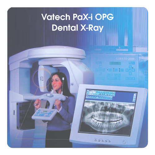
An OPG (Orthopantomagram) is an all panoramic examining dental X-ray of the upper and lower jaw. It is likewise some of the time called Orthopantomagraph or by the restrictive name Panorex. It shows a straightened two-dimensional perspective on a half-circle from one ear to another. All-encompassing x-beams permit pictures of different points to be taken to make up the composite all-encompassing picture, where the maxilla (upper jaw) and mandible (lower jaw) are in the seen region. The constructions that are outside the seen region are obscured. At some stage in your dental treatment, your dental specialist will probably take an OPG.
An OPG in like manner shows the number, position, and improvement of the large number of teeth including those that needy individuals yet surfaced or shot out through the gum. It is not quite the same as the little close-up x-ray dental specialists take of individual teeth. It shows less fine detail, however a lot more extensive space of view. This can be especially valuable to check hard-to-see regions like insight teeth, or the improvement of a youngster’s jaw and teeth, helpful for surveying for advancement for the most part, yet in addition to orthodontic need. It is additionally normally used to check your jaw joint, the TMJ (temporomandibular joint), now and then called the CMA (craniomandibular verbalization), particularly assuming you grate your teeth.
The main benefit of all-encompassing pictures/OPGs:
Wide inclusion of facial bones and teeth including the TMJ (temporomandibular joint)
Low quiet radiation portion
The convenience of examination for the patient
Ability to be utilized in patients who are confined in opening their mouth
Brief time frame needed for producing the picture.
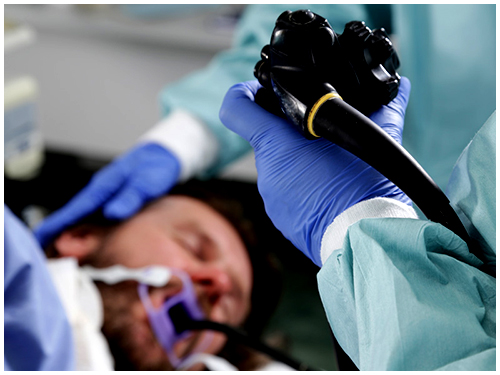
An endoscopy is a procedure used in medicine to look inside the body. The endoscopy procedure uses an endoscope to examine the interior of a hollow organ or cavity of the body. Unlike many other medical imaging techniques, endoscopes are inserted directly into the organ.
There are many types of endoscopes. Depending on the site in the body and type of procedure an endoscopy may be performed either by a doctor or a surgeon. A patient may be fully conscious or anaesthetised during the procedure.
There are several types of endoscopy.
Tesophagogastroduodenoscopy (EGD) which is often called upper endoscopy, gastroscopy, enteroscopy, endoscopic ultrasound (EUS), endoscopic retrograde cholangiopancreatography (ERCP), colonoscopy, and sigmoidoscopy. Percutaneous endoscopic gastrostomy (PEG) is a procedure that utilizes endoscopy to help
placement of a tube into the stomach; a small incision in the skin is also required.
 A Home Blood Draw Service brings the convenience of medical testing to your doorstep. Instead of going to a medical facility, you can have a trained phlebotomist visit your home to collect blood samples for various tests. This service is especially beneficial for individuals who have difficulty traveling or prefer the comfort and privacy of their own homes.
By opting for a Home Blood Draw Service, you can avoid the long waits, crowded waiting areas, and the inconvenience of taking time off work or arranging transportation to visit a medical facility. This service is designed to cater to your needs and provide you with a hassle-free experience while ensuring accurate and reliable blood test results.
- Convenience: The primary benefit of a Home Blood Draw Service is the convenience it offers. You no longer have to rearrange your schedule or face the stress of commuting to a medical facility for a blood test. Instead, a phlebotomist will come to your home at a time that is convenient for you, eliminating the need for unnecessary travel and waiting.
- Comfort: Getting your blood drawn in the comfort of your own home can make the experience much more comfortable and less intimidating. You can relax in a familiar environment, which can help reduce anxiety and make the process more pleasant for individuals who may feel uneasy or have a fear of medical settings.
- Personalized and Individualized Care: When you choose a Home Blood Draw Service, you can expect personalized care tailored to your specific needs. The phlebotomist will take the time to understand your requirements and address any concerns you may have. This individualized approach ensures that you receive the attention and care you deserve, leading to a better overall experience.
- Time-Saving: With a Home Blood Draw Service, you can save valuable time that would otherwise be spent traveling to and from a medical facility and waiting for your turn. This is particularly beneficial for individuals with busy schedules or those who have difficulty leaving their homes due to mobility issues or chronic conditions.
- Reduced Risk of Exposure: In today’s world, where infectious diseases are a concern, a Home Blood Draw Service provides an added layer of safety by minimizing your exposure to crowded waiting areas and other potentially contaminated surfaces. By staying in the comfort of your home, you can reduce the risk of contracting illnesses and ensure your safety.
- Access to Specialized Testing: Some home blood draw service providers offer specialized testing options that may not be readily available at all medical facilities. This can be particularly beneficial for individuals with rare conditions or those who require specific tests that are not commonly offered in traditional settings.